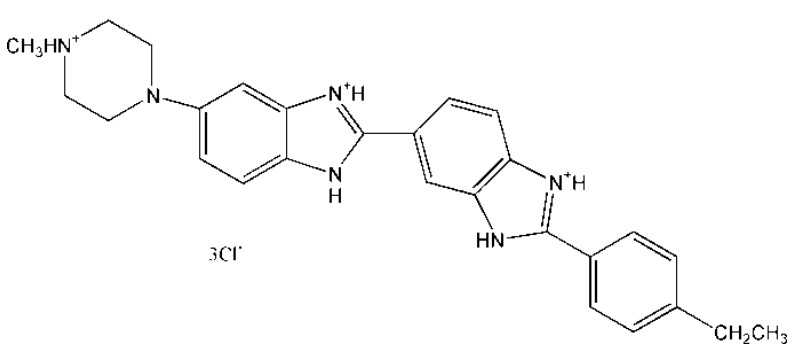
Hoechst33342是一种可以穿透细胞膜的蓝色荧光染料,它在嵌入双链DNA后释放强烈的蓝色荧光,对细胞的毒性较低。Hoechst33342常用于细胞凋亡检测,染色后用荧光显微镜观察或流式细胞仪检测。也可以用于普通的细胞核染色,或常规的DNA染色。
本品为粉末状,用于细胞核染色时,推荐的Hoechst33342工作浓度为0.5-10μg/mL。如需特定浓度或者经浓度优化的产品,可选购Hoechst33342染液(1mg/ml)(CatNo.40732ES03)
| 中文名称(Chinese Synonym) | 二苯甲亚胺, 三氯化氢 2'-(4-乙基苯基)-5-(4-甲基-1-哌嗪基)-2,5'-二-1H-苯并咪唑 |
| 英文名称(English Synonym) | 2’-(4-Ethoxyphenyl)-5-(4-methyl-1-piperazinyl)-2,5’-bi-1H-benzimidazole, trihydrochloride |
| 分子式(Molecular Formula) | C27H28N6O·3HCl |
| 分子量(Molecular Weight) | 561.93 |
| CAS号(CAS NO.) | 23491-52-3 |
| 荧光光谱(Fluorescence Spectral) | Hoechst 33342的Ex/Em=346/460; Hoechst33342-核酸的Ex/Em=350/461. 荧光可用氙气灯,泵弧灯或者UV激光激发;可用DAPI滤光器、蓝色GFP滤光器、Alexa Fluor 350滤光器等检测。 |
| 纯度(Purity) | ≥95%(HPLC) |
| 结构(Structure) |  |
冰袋(wet ice)运输。-20℃干燥避光保存,有效期2年。
[1] Zhai Y, Wang J, Lang T, et al. T lymphocyte membrane-decorated epigenetic nanoinducer of interferons for cancer immunotherapy. Nat Nanotechnol. 2021;16(11):1271-1280. doi:10.1038/s41565-021-00972-7(IF:39.213)
[2] Zhang W, Liu J, Li X, et al. Precise Chemodynamic Therapy of Cancer by Trifunctional Bacterium-Based Nanozymes. ACS Nano. 2021;15(12):19321-19333. doi:10.1021/acsnano.1c05605(IF:15.881)
[3] Yu H, Fan J, Shehla N, et al. Biomimetic Hybrid Membrane-Coated Xuetongsu Assisted with Laser Irradiation for Efficient Rheumatoid Arthritis Therapy [published online ahead of print, 2021 Dec 29]. ACS Nano. 2021;10.1021/acsnano.1c07556. doi:10.1021/acsnano.1c07556(IF:15.881)
[4] Wang Z, Gong X, Li J, et al. Oxygen-Delivering Polyfluorocarbon Nanovehicles Improve Tumor Oxygenation and Potentiate Photodynamic-Mediated Antitumor Immunity. ACS Nano. 2021;15(3):5405-5419. doi:10.1021/acsnano.1c00033(IF:15.881)
[5] Wang D, Dong H, Li M, et al. Erythrocyte-Cancer Hybrid Membrane Camouflaged Hollow Copper Sulfide Nanoparticles for Prolonged Circulation Life and Homotypic-Targeting Photothermal/Chemotherapy of Melanoma. ACS Nano. 2018;12(6):5241-5252. doi:10.1021/acsnano.7b08355(IF:13.709)
[6] Ma M, Chen Y, Zhao M, et al. Hierarchical responsive micelle facilitates intratumoral penetration by acid-activated positive charge surface and size contraction. Biomaterials. 2021;271:120741. doi:10.1016/j.biomaterials.2021.120741(IF:12.479)
[7] Wang Q, Duan X, Huang F, et al. Polystyrene nanoplastics alter virus replication in orange-spotted grouper (Epinephelus coioides) spleen and brain tissues and spleen cells. J Hazard Mater. 2021;416:125918. doi:10.1016/j.jhazmat.2021.125918(IF:10.588)
[8] Zhao Q, Jiang D, Sun X, et al. Biomimetic nanotherapy: core-shell structured nanocomplexes based on the neutrophil membrane for targeted therapy of lymphoma. J Nanobiotechnology. 2021;19(1):179. Published 2021 Jun 13. doi:10.1186/s12951-021-00922-4(IF:10.435)
[9] Huang R, Cai GQ, Li J, et al. Platelet membrane-camouflaged silver metal-organic framework drug system against infections caused by methicillin-resistant Staphylococcus aureus [published correction appears in J Nanobiotechnology. 2021 Sep 19;19(1):278]. J Nanobiotechnology. 2021;19(1):229. Published 2021 Aug 4. doi:10.1186/s12951-021-00978-2(IF:10.435)
[10] Qi N, Zhang S, Zhou X, et al. Combined integrin αvβ3 and lactoferrin receptor targeted docetaxel liposomes enhance the brain targeting effect and anti-glioma effect. J Nanobiotechnology. 2021;19(1):446. Published 2021 Dec 23. doi:10.1186/s12951-021-01180-0(IF:10.435)
[11] Wang H, Li J, Wang Z, et al. Tumor-permeated bioinspired theranostic nanovehicle remodels tumor immunosuppression for cancer therapy. Biomaterials. 2021;269:120609. doi:10.1016/j.biomaterials.2020.120609(IF:10.317)
[12] Han C, Xu X, Zhang C, et al. Cytochrome c light-up graphene oxide nanosensor for the targeted self-monitoring of mitochondria-mediated tumor cell death [published online ahead of print, 2020 Nov 5]. Biosens Bioelectron. 2020;173:112791. doi:10.1016/j.bios.2020.112791(IF:10.257)
[13] Yang Q, Yang F, Dai W, et al. DNA Logic Circuits for Multiple Tumor Cells Identification Using Intracellular MicroRNA Molecular Bispecific Recognition. Adv Healthc Mater. 2021;10(21):e2101130. doi:10.1002/adhm.202101130(IF:9.933)
[14] Lin Y, Yi O, Hu M, et al. Multifunctional nanoparticles of sinomenine hydrochloride for treat-to-target therapy of rheumatoid arthritis via modulation of proinflammatory cytokines [published online ahead of print, 2022 Jun 2]. J Control Release. 2022;348:42-56. doi:10.1016/j.jconrel.2022.05.016(IF:9.776)
[15] Yu M, Yu J, Yi Y, et al. Oxidative stress-amplified nanomedicine for intensified ferroptosis-apoptosis combined tumor therapy. J Control Release. 2022;347:104-114. doi:10.1016/j.jconrel.2022.04.047(IF:9.776)
[16] Dai W, Su L, Lu H, Dong H, Zhang X. Exosomes-mediated synthetic Dicer substrates delivery for intracellular Dicer imaging detection. Biosens Bioelectron. 2020;151:111907. doi:10.1016/j.bios.2019.111907(IF:9.518)
[17] Sun Y, Zhou Y, Shi Y, et al. Expression of miRNA-29 in Pancreatic β Cells Promotes Inflammation and Diabetes via TRAF3. Cell Rep. 2021;34(1):108576. doi:10.1016/j.celrep.2020.108576(IF:9.423)
[18] Zhao Q, Li J, Wu B, et al. Smart Biomimetic Nanocomposites Mediate Mitochondrial Outcome through Aerobic Glycolysis Reprogramming: A Promising Treatment for Lymphoma. ACS Appl Mater Interfaces. 2020;12(20):22687-22701. doi:10.1021/acsami.0c05763(IF:9.229)
[19] Li X, Gui R, Li J, et al. Novel Multifunctional Silver Nanocomposite Serves as a Resistance-Reversal Agent to Synergistically Combat Carbapenem-Resistant Acinetobacter baumannii. ACS Appl Mater Interfaces. 2021;13(26):30434-30457. doi:10.1021/acsami.1c10309(IF:9.229)
[20] Xu H, Liao C, Liang S, Ye BC. A Novel Peptide-Equipped Exosomes Platform for Delivery of Antisense Oligonucleotides. ACS Appl Mater Interfaces. 2021;13(9):10760-10767. doi:10.1021/acsami.1c00016(IF:9.229)
[21] Luo Q, Lin L, Huang Q, et al. Dual stimuli-responsive dendronized prodrug derived from poly(oligo-(ethylene glycol) methacrylate)-based copolymers for enhanced anti-cancer therapeutic effect. Acta Biomater. 2022;143:320-332. doi:10.1016/j.actbio.2022.02.033(IF:8.947)
[22] Su J, Sun H, Meng Q, Zhang P, Yin Q, Li Y. Enhanced Blood Suspensibility and Laser-Activated Tumor-specific Drug Release of Theranostic Mesoporous Silica Nanoparticles by Functionalizing with Erythrocyte Membranes [published correction appears in Theranostics. 2020 Jan 18;10(5):2401]. Theranostics. 2017;7(3):523-537. Published 2017 Jan 7. doi:10.7150/thno.17259(IF:8.766)
[23] Kang S, Duan W, Zhang S, Chen D, Feng J, Qi N. Muscone/RI7217 co-modified upward messenger DTX liposomes enhanced permeability of blood-brain barrier and targeting glioma. Theranostics. 2020;10(10):4308-4322. Published 2020 Mar 4. doi:10.7150/thno.41322(IF:8.579)
[24] Cao Y, Li S, Chen C, et al. Rattle-type Au@Cu2-xS hollow mesoporous nanocrystals with enhanced photothermal efficiency for intracellular oncogenic microRNA detection and chemo-photothermal therapy. Biomaterials. 2018;158:23-33. doi:10.1016/j.biomaterials.2017.12.009(IF:8.402)
[25] Chen P, Kuang W, Zheng Z, et al. Carboxylesterase-Cleavable Biotinylated Nanoparticle for Tumor-Dual Targeted Imaging. Theranostics. 2019;9(24):7359-7369. Published 2019 Sep 25. doi:10.7150/thno.37625(IF:8.063)
[26] Yu J, Liu S, Chen L, Wu B. Combined effects of arsenic and palmitic acid on oxidative stress and lipid metabolism disorder in human hepatoma HepG2 cells. Sci Total Environ. 2021;769:144849. doi:10.1016/j.scitotenv.2020.144849(IF:7.963)
[27] Yang X, Shi X, Zhang Y, et al. Photo-triggered self-destructive ROS-responsive nanoparticles of high paclitaxel/chlorin e6 co-loading capacity for synergetic chemo-photodynamic therapy. J Control Release. 2020;323:333-349. doi:10.1016/j.jconrel.2020.04.027(IF:7.727)
[28] Ji Y, Sun Y, Hei M, et al. NIR Activated Upper Critical Solution Temperature Polymeric Micelles for Trimodal Combinational Cancer Therapy. Biomacromolecules. 2022;23(3):937-947. doi:10.1021/acs.biomac.1c01356(IF:6.988)
[29] Cheng Y , Lu H , Yang F , Zhang Y , Dong H . Biodegradable FeWOx nanoparticles for CT/MR imaging-guided synergistic photothermal, photodynamic, and chemodynamic therapy. Nanoscale. 2021;13(5):3049-3060. doi:10.1039/d0nr07215j(IF:6.895)
[30] Gao YR, Zhang WX, Wei YN, et al. Ionic liquids enable the preparation of a copper-loaded gel with transdermal delivery function for wound dressings. Biomater Sci. 2022;10(4):1041-1052. Published 2022 Feb 15. doi:10.1039/d1bm01745d(IF:6.843)
[31] Xie Q, Liu Y, Long Y, et al. Hybrid-cell membrane-coated nanocomplex-loaded chikusetsusaponin IVa methyl ester for a combinational therapy against breast cancer assisted by Ce6. Biomater Sci. 2021;9(8):2991-3004. doi:10.1039/d0bm02211j(IF:6.843)
[32] Yan P, Shu X, Zhong H, et al. A versatile nanoagent for multimodal imaging-guided photothermal and anti-inflammatory combination cancer therapy. Biomater Sci. 2021;9(14):5025-5034. doi:10.1039/d1bm00576f(IF:6.843)
[33] Wang Q, Bu S, Xin D, Li B, Wang L, Lai D. Autophagy Is Indispensable for the Self-Renewal and Quiescence of Ovarian Cancer Spheroid Cells with Stem Cell-Like Properties. Oxid Med Cell Longev. 2018;2018:7010472. Published 2018 Sep 17. doi:10.1155/2018/7010472(IF:6.543)
[34] Huang Q, Gao W, Mu H, et al. HSP60 Regulates Monosodium Urate Crystal-Induced Inflammation by Activating the TLR4-NF-κB-MyD88 Signaling Pathway and Disrupting Mitochondrial Function. Oxid Med Cell Longev. 2020;2020:8706898. Published 2020 Dec 31. doi:10.1155/2020/8706898(IF:6.543)
[35] Liu S, Jiang W, Wu B, et al. Low levels of graphene and graphene oxide inhibit cellular xenobiotic defense system mediated by efflux transporters. Nanotoxicology. 2016;10(5):597-606. doi:10.3109/17435390.2015.1104739(IF:6.411)
[36] Long Y, Wang Z, Fan J, et al. A hybrid membrane coating nanodrug system against gastric cancer via the VEGFR2/STAT3 signaling pathway. J Mater Chem B. 2021;9(18):3838-3855. doi:10.1039/d1tb00029b(IF:6.331)
[37] Guo W, Ma X, Fu Y, et al. Discovering and Characterizing of Survivin Dominant Negative Mutants With Stronger Pro-apoptotic Activity on Cancer Cells and CSCs. Front Oncol. 2021;11:635233. Published 2021 Mar 31. doi:10.3389/fonc.2021.635233(IF:6.244)
[38] Sun Y, Zhou S, Shi Y, et al. Inhibition of miR-153, an IL-1β-responsive miRNA, prevents beta cell failure and inflammation-associated diabetes. Metabolism. 2020;111:154335. doi:10.1016/j.metabol.2020.154335(IF:6.159)
[39] Wang R, Yang H, Fu R, et al. Biomimetic Upconversion Nanoparticles and Gold Nanoparticles for Novel Simultaneous Dual-Modal Imaging-Guided Photothermal Therapy of Cancer. Cancers (Basel). 2020;12(11):3136. Published 2020 Oct 27. doi:10.3390/cancers12113136(IF:6.126)
[40] Daniyal M, Liu Y, Yang Y, et al. Anti-gastric cancer activity and mechanism of natural compound "Heilaohulignan C" isolated from Kadsura coccinea. Phytother Res. 2021;35(7):3977-3987. doi:10.1002/ptr.7114(IF:5.882)
[41] Zhu S, Liu F, Zeng H, et al. Insulin/IGF signaling and TORC1 promote vitellogenesis via inducing juvenile hormone biosynthesis in the American cockroach. Development. 2020;147(20):dev188805. Published 2020 Oct 23. doi:10.1242/dev.188805(IF:5.611)
[42] Wang J, Nisar M, Huang C, et al. Small molecule natural compound agonist of SIRT3 as a therapeutic target for the treatment of intervertebral disc degeneration. Exp Mol Med. 2018;50(11):1-14. Published 2018 Nov 12. doi:10.1038/s12276-018-0173-3(IF:5.584)
[43] Yu J, Liu S, Wu B, et al. Comparison of Cytotoxicity and Inhibition of Membrane ABC Transporters Induced by MWCNTs with Different Length and Functional Groups. Environ Sci Technol. 2016;50(7):3985-3994. doi:10.1021/acs.est.5b05772(IF:5.393)
[44] Li W, Xie X, Wu T, et al. Loading Auristatin PE onto boron nitride nanotubes and their effects on the apoptosis of Hep G2 cells. Colloids Surf B Biointerfaces. 2019;181:305-314. doi:10.1016/j.colsurfb.2019.05.047(IF:5.268)
[45] Hussain M, Hamid MI, Wang N, Bin L, Xiang M, Liu X. The transcription factor SKN7 regulates conidiation, thermotolerance, apoptotic-like cell death and parasitism in the nematode endoparasitic fungus Hirsutella minnesotensis. Sci Rep. 2016;6:30047. Published 2016 Jul 20. doi:10.1038/srep30047(IF:5.228)
[46] Wang F, Xiao F, Du L, et al. Activation of GCN2 in macrophages promotes white adipose tissue browning and lipolysis under leucine deprivation. FASEB J. 2021;35(6):e21652. doi:10.1096/fj.202100061RR(IF:5.192)
[47] Zhang C, Lei JL, Zhang H, et al. Calyxin Y sensitizes cisplatin-sensitive and resistant hepatocellular carcinoma cells to cisplatin through apoptotic and autophagic cell death via SCF βTrCP-mediated eEF2K degradation. Oncotarget. 2017;8(41):70595-70616. Published 2017 Aug 3. doi:10.18632/oncotarget.19883(IF:5.168)
[48] Yu P, Zhang C, Gao CY, et al. Anti-proliferation of triple-negative breast cancer cells with physagulide P: ROS/JNK signaling pathway induces apoptosis and autophagic cell death. Oncotarget. 2017;8(38):64032-64049. Published 2017 Jul 17. doi:10.18632/oncotarget.19299(IF:5.168)
[49] Wang Y, Liu Y, Wu B, Rui M, Liu J, Lu G. Comparison of toxicity induced by EDTA-Cu after UV/H2O2 and UV/persulfate treatment: Species-specific and technology-dependent toxicity. Chemosphere. 2020;240:124942. doi:10.1016/j.chemosphere.2019.124942(IF:5.108)
[50] Wu B, Wu X, Liu S, Wang Z, Chen L. Size-dependent effects of polystyrene microplastics on cytotoxicity and efflux pump inhibition in human Caco-2 cells. Chemosphere. 2019;221:333-341. doi:10.1016/j.chemosphere.2019.01.056(IF:5.108)
[51] Wang D, Liu Y, Zhang R, et al. Apoptotic transition of senescent cells accompanied with mitochondrial hyper-function. Oncotarget. 2016;7(19):28286-28300. doi:10.18632/oncotarget.8536(IF:5.008)
[52] Dang W, Liu H, Fan J, et al. Monitoring VEGF mRNA and imaging in living cells in vitro using rGO-based dual fluorescent signal amplification platform. Talanta. 2019;205:120092. doi:10.1016/j.talanta.2019.06.092(IF:4.916)
[53] Dang W, Luo R, Fan J, et al. RNase A activity analysis and imaging using label-free DNA-templated silver nanoclusters. Talanta. 2020;209:120512. doi:10.1016/j.talanta.2019.120512(IF:4.916)
[54] Chen G, Kong Y, Li Y, et al. A Promising Intracellular Protein-Degradation Strategy: TRIMbody-Away Technique Based on Nanobody Fragment. Biomolecules. 2021;11(10):1512. Published 2021 Oct 14. doi:10.3390/biom11101512(IF:4.879)
[55] Yuan J, Li X, Zhang G, et al. USP39 mediates p21-dependent proliferation and neoplasia of colon cancer cells by regulating the p53/p21/CDC2/cyclin B1 axis. Mol Carcinog. 2021;60(4):265-278. doi:10.1002/mc.23290(IF:4.784)
[56] Xiao L, Ding Z, Zhang X, Wang X, Lu Q, Kaplan DL. Silk Nanocarrier Size Optimization for Enhanced Tumor Cell Penetration and Cytotoxicity In Vitro. ACS Biomater Sci Eng. 2022;8(1):140-150. doi:10.1021/acsbiomaterials.1c01122(IF:4.749)
[57] Huang H, Zou X, Zhong L, et al. CRISPR/dCas9-mediated activation of multiple endogenous target genes directly converts human foreskin fibroblasts into Leydig-like cells. J Cell Mol Med. 2019;23(9):6072-6084. doi:10.1111/jcmm.14470(IF:4.658)
[58] Huang H, Zhong L, Zhou J, et al. Leydig-like cells derived from reprogrammed human foreskin fibroblasts by CRISPR/dCas9 increase the level of serum testosterone in castrated male rats. J Cell Mol Med. 2020;24(7):3971-3981. doi:10.1111/jcmm.15018(IF:4.486)
[59] Huang X, Wu B, Li J, et al. Anti-tumour effects of red blood cell membrane-camouflaged black phosphorous quantum dots combined with chemotherapy and anti-inflammatory therapy. Artif Cells Nanomed Biotechnol. 2019;47(1):968-979. doi:10.1080/21691401.2019.1584110(IF:4.462)
[60] Huang K, Boerhan R, Liu C, Jiang G. Nanoparticles Penetrate into the Multicellular Spheroid-on-Chip: Effect of Surface Charge, Protein Corona, and Exterior Flow. Mol Pharm. 2017;14(12):4618-4627. doi:10.1021/acs.molpharmaceut.7b00726(IF:4.440)
[61] Liu M, Fu M, Yang X, et al. Paclitaxel and quercetin co-loaded functional mesoporous silica nanoparticles overcoming multidrug resistance in breast cancer. Colloids Surf B Biointerfaces. 2020;196:111284. doi:10.1016/j.colsurfb.2020.111284(IF:4.389)
[62] Zhang Q, Wu L, Liu S, et al. Targeted nanobody complex enhanced photodynamic therapy for lung cancer by overcoming tumor microenvironment. Cancer Cell Int. 2020;20(1):570. Published 2020 Nov 27. doi:10.1186/s12935-020-01613-0(IF:4.175)
[63] Xu YN, Wang Z, Zhang SK, et al. Low-grade elevation of palmitate and lipopolysaccharide synergistically induced β-cell damage via inhibition of neutral ceramidase. Mol Cell Endocrinol. 2022;539:111473. doi:10.1016/j.mce.2021.111473(IF:4.102)
[64] Liu J, Li C, Cheng S, Ya S, Gao D, Ding W. Large-scale high-density culture of hepatocytes in a liver microsystem with mimicked sinusoid blood flow. J Tissue Eng Regen Med. 2018;12(12):2266-2276. doi:10.1002/term.2758(IF:4.089)
[65] Hui Y, Tang T, Wang J, et al. Fusaricide is a Novel Iron Chelator that Induces Apoptosis through Activating Caspase-3. J Nat Prod. 2021;84(8):2094-2103. doi:10.1021/acs.jnatprod.0c01322(IF:4.050)
[66] Zhao M , Gao Y , Ye S , et al. A light-up near-infrared probe with aggregation-induced emission characteristics for highly sensitive detection of alkaline phosphatase. Analyst. 2019;144(21):6262-6269. doi:10.1039/c9an01505a(IF:4.019)
[67] Shen Z, Wu J, Yu Y, et al. Comparison of cytotoxicity and membrane efflux pump inhibition in HepG2 cells induced by single-walled carbon nanotubes with different length and functional groups. Sci Rep. 2019;9(1):7557. Published 2019 May 17. doi:10.1038/s41598-019-43900-5(IF:4.011)
[68] Yu Y, Wu B, Jiang L, Zhang XX, Ren HQ, Li M. Comparative analysis of toxicity reduction of wastewater in twelve industrial park wastewater treatment plants based on battery of toxicity assays. Sci Rep. 2019;9(1):3751. Published 2019 Mar 6. doi:10.1038/s41598-019-40154-z(IF:4.011)
[69] Wang Y, Cui S, Wu B, Zhang Q, Jiang W. PLA-based core-shell structure stereocomplexed nanoparticles with enhanced loading and release profile of paclitaxel. Front Biosci (Landmark Ed). 2021;26(9):517-532. doi:10.52586/4964(IF:4.009)
[70] Lu G, Qin D, Wang Y, Liu J, Chen W. Single and combined effects of selected haloacetonitriles in a human-derived hepatoma line. Ecotoxicol Environ Saf. 2018;163:417-426. doi:10.1016/j.ecoenv.2018.07.104(IF:3.974)
[71] Shang Y, Wang Q, Li J, et al. Platelet-Membrane-Camouflaged Zirconia Nanoparticles Inhibit the Invasion and Metastasis of Hela Cells. Front Chem. 2020;8:377. Published 2020 May 7. doi:10.3389/fchem.2020.00377(IF:3.693)
[72] Shi H, Mao Y, Ju Q, et al. C-terminal binding protein‑2 mediates cisplatin chemoresistance in esophageal cancer cells via the inhibition of apoptosis. Int J Oncol. 2018;53(1):167-176. doi:10.3892/ijo.2018.4367(IF:3.333)
[73] Wu W, Wu Q, Liu X. Chronic activation of FXR-induced liver growth with tissue-specific targeting Cyclin D1. Cell Cycle. 2019;18(15):1784-1797. doi:10.1080/15384101.2019.1634955(IF:3.259)
[74] Zhu D, Chen C, Xia Y, Kong LY, Luo J. A Purified Resin Glycoside Fraction from Pharbitidis Semen Induces Paraptosis by Activating Chloride Intracellular Channel-1 in Human Colon Cancer Cells. Integr Cancer Ther. 2019;18:1534735418822120. doi:10.1177/1534735418822120(IF:2.634)
[75] Wang Y, Liu Y, Liu S, Wu B. Influence of Iron on Cytotoxicity and Gene Expression Profiles Induced by Arsenic in HepG2 Cells. Int J Environ Res Public Health. 2019;16(22):4484. Published 2019 Nov 14. doi:10.3390/ijerph16224484(IF:2.468)
[76] Song X, Tang Y, Zhu J, et al. HIF-1α induces hypoxic apoptosis of MLO-Y4 osteocytes via JNK/caspase-3 pathway and the apoptotic-osteocyte-mediated osteoclastogenesis in vitro. Tissue Cell. 2020;67:101402. doi:10.1016/j.tice.2020.101402(IF:2.466)
[77] Du J, Chen C, Sun Y, Zheng L, Wang W. Ponicidin suppresses HT29 cell growth via the induction of G1 cell cycle arrest and apoptosis. Mol Med Rep. 2015;12(4):5816-5820. doi:10.3892/mmr.2015.4150(IF:1.554)


