细胞增殖检测方法大全(下)——荧光染料法
细胞增殖是生物体生长、发育、繁殖和遗传的基础,是生物体的重要生命特征。细胞通常以分裂的方式进行增殖。细胞毒性(cytotoxic)是由细胞或化学物质引起的单纯细胞杀伤事件,不依赖于凋亡或坏死的细胞死亡机理。
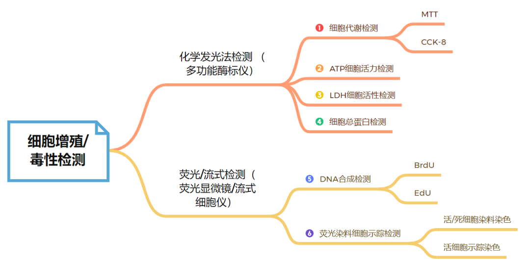
根据您实验室现有的实验条件,可选择使用多功能酶标仪的化学发光发检测,也可选择利用荧光显微镜或者流式细胞仪的检测。最常见的细胞增殖和毒性的检测方法有以下几种:
原理解读

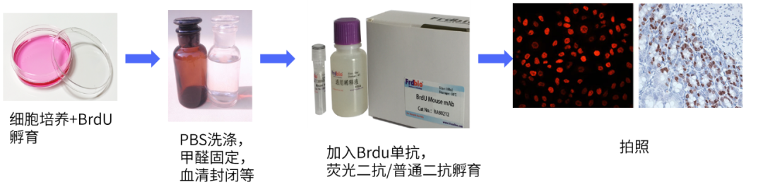
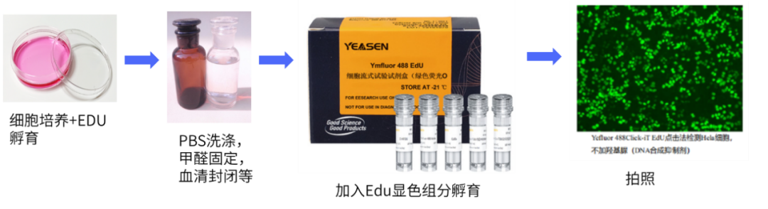
实验操作
实验结果
-
荧光图片结果:
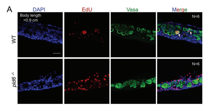
参考文献:Zhang R, Tu YX, Ye D, Gu Z, Chen ZX, Sun Y. A Germline-Specific Regulator of Mitochondrial Fusion is Required for Maintenance and Differentiation of Germline Stem and Progenitor Cells [published online ahead of print, 2022 Oct 18]. Adv Sci (Weinh). 2022;e2203631. doi:10.1002/advs.202203631(IF:17.521)
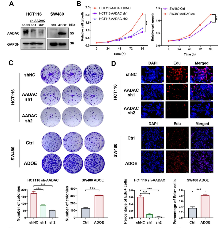
参考文献:Sun R, Lin Z, Wang X, et al. AADAC protects colorectal cancer liver colonization from ferroptosis through SLC7A11-dependent inhibition of lipid peroxidation [published correction appears in J Exp Clin Cancer Res. 2022 Oct 25;41(1):313]. J Exp Clin Cancer Res. 2022;41(1):284. Published 2022 Sep 26. doi:10.1186/s13046-022-02493-0(IF:12.658)
-
流式图片结果:
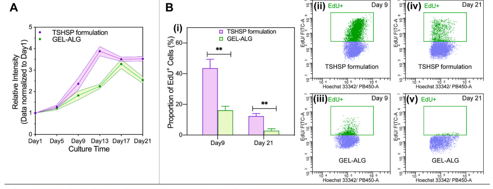
参考文献:Chen H, Fei F, Li X, et al. A structure-supporting, self-healing, and high permeating hydrogel bioink for establishment of diverse homogeneous tissue-like constructs. Bioact Mater. 2021;6(10):3580-3595. Published 2021 Mar 23. doi:10.1016/j.bioactmat.2021.03.019(IF:14.593)
|
货号 |
产品名称 |
规格 |
|
40275ES60/76/80 |
100/500/1000T |
|
|
40276ES60/76/80 |
100/500/1000T |
|
|
40277ES60/76/80 |
100/500/1000T |
|
|
40278ES25/50 |
Yefluor 488 EdU Flow Cytometry Assay Kits |
20/50T |
|
40279ES25/50 |
Yefluor 594 EdU Flow Cytometry Assay Kits |
20/50T |
|
40280ES25/50 |
Yefluor 647 EdU Flow Cytometry Assay Kits |
20/50T |
|
产品货号 |
产品名称 |
Ex/Em nm |
染色部位 |
应用 |
|
40757ES25 |
748/780 |
细胞膜染色,深红色荧光 |
活细胞染色,体内成像或示踪实验,外泌体染色 |
|
|
40725ES10/25 |
484/501 |
细胞膜染色,绿色荧光 |
活细胞染色,体内成蜃 |
|
|
40758ES25 |
644/663 |
细胞膜染色,红色荧光 |
外泌体染色,活细胞染色,自带其他荧光的细胞或组织染色 |
|
|
40718ES50/60/72 |
553/570 |
细胞膜染色,红色荧光 |
活体动物染色,细胞染色 |
|
|
40714ES76/80 |
CFDA SE Cell Proliferation and Cell Tracking Kit |
494/521 |
细胞浆染色,绿色荧光 |
细胞增殖群落分析,体内增殖研究 |
|
40721ES50/60/72 |
492/517 |
细胞浆染色,绿色荧光 |
细胞增殖,细胞间融合,细胞粘附及多药耐药转运蛋白的研究,病原菌标记 |
|
|
40747ES76/80 |
490/515 |
Calcein-AM:活细胞浆染色,发绿色荧光;P1:死细胞核染色,红色荧光 |
活细胞/死细胞染色 |
|
|
40711ES10/60 |
535/617 |
细胞核内DNA |
死细胞染色 |
|
|
40745ES64 |
546/647 |
细胞核内DNA |
坏死或晚期凋亡细胞染色 |
|
|
40202ES60/76/80/92 |
560/590 |
在氧化状态下呈现紫蓝色无荧光性,而在细胞增殖旺盛期,还原状态下,转变为呈粉红或红色荧光的还原产物 |
动物、真菌和细菌细胞的增殖情况 |
CFDA,SE染料检测细胞增殖与示踪
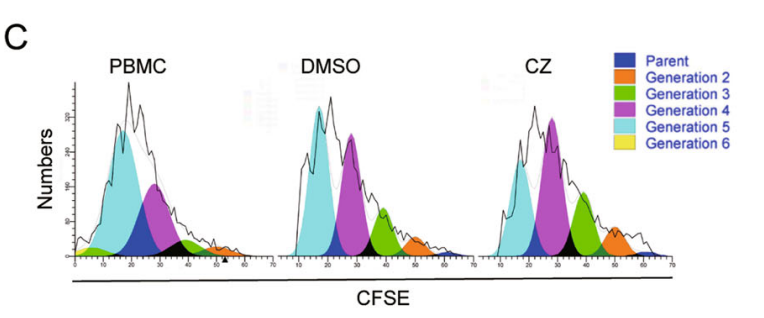
参考文献:Deng, Luchan et al. “Chlorzoxazone, a small molecule drug, augments immunosuppressive capacity of mesenchymal stem cells via modulation of FOXO3 phosphorylation.” Cell death & disease vol. 11,3 158. 2 Mar. 2020, doi:10.1038/s41419-020-2357-8
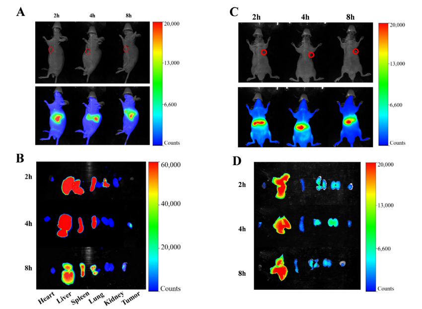
DiR染料标记肿瘤细胞后,注射肿瘤细胞进入动物体内,活体成像
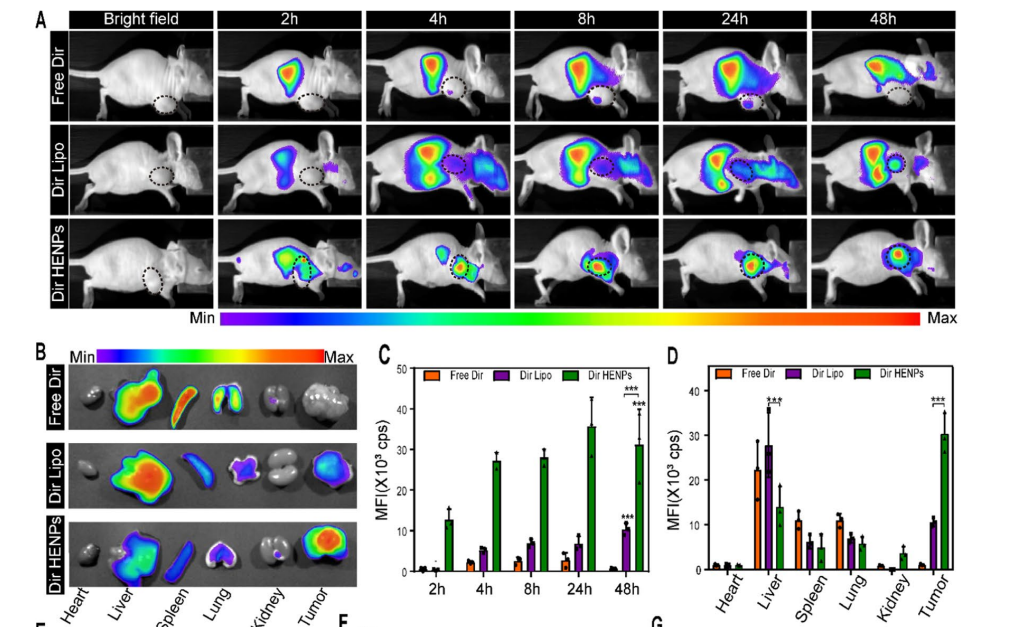
参考文献:Li L, He D, Guo Q, et al. Exosome-liposome hybrid nanoparticle codelivery of TP and miR497 conspicuously overcomes chemoresistant ovarian cancer. J Nanobiotechnology. 2022;20(1):50. Published 2022 Jan 25. doi:10.1186/s12951-022-01264-5(IF:10.435)
DiR染料标记外泌体后,注射外泌体进入动物体内,活体成像
Calcein-AM/PI活细胞/死细胞染色
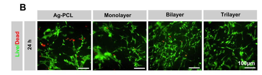
参考文献:Zhang W, Xia S, Weng T, et al. Antibacterial coaxial hydro-membranes accelerate diabetic wound healing by tuning surface immunomodulatory functions. Mater Today Bio. 2022;16:100395. Published 2022 Aug 13. doi:10.1016/j.mtbio.2022.100395(IF:10.761)
阿尔玛蓝染色
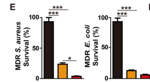
参考文献:Yin H, Zhou M, Chen X, et al. Fructose-coated Ångstrom silver prevents sepsis by killing bacteria and attenuating bacterial toxin-induced injuries. Theranostics. 2021;11(17):8152-8171. Published 2021 Jul 13. doi:10.7150/thno.55334(IF:11.556)
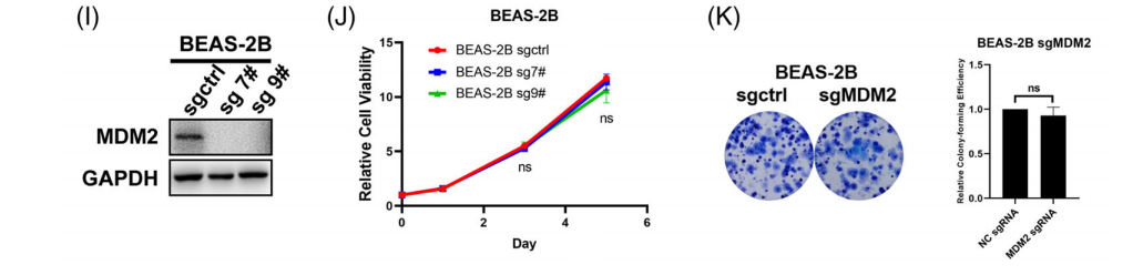
参考文献:Wang Q, Li J, Zhu J, et al. Genome-wide CRISPR/Cas9 screening for therapeutic targets in NSCLC carrying wild-type TP53 and receptor tyrosine kinase genes. Clin Transl Med. 2022;12(6):e882. doi:10.1002/ctm2.882(IF:11.492)
染色原理
|
货号 |
产品名称 |
规格 |
|
40207ES20 |
20mL |
|
|
40207ES60 |
100mL |
|
|
40208ES60 |
Typan Blue Staining Cell Viability Assay Kit |
100T |
|
40208ES76 |
500T |