如何完成一次高效又经济的Tunel检测?
——Yeasen TUNEL detection Kit 让“美”原位呈现
通过TdT转移酶将Fluorescein-dUTP结合到有缺口的DNA上是检测细胞凋亡的常用方法之一,如Roche提供的In Situ Cell Death Kit正是基于这个原理。产品中的TdT转移酶活性直接影响到标记的效率。翌圣生物立足于酶制剂生产,力求开发以酶制剂为基础的较高具性价比产品,推出三款荧光素(Alexa Fluor 640,Alexa Fluor 488,FITC)标记的Tunel检测试剂,在满足客户对产品高品质要求的同时又可以享受低至Roche一半的价格。
目前已经有超过120个课题组正在使用该产品,并在权威杂志上发表多篇英文文章,如Theranostics和EMBO Molecular Medicine等。
检测机理:
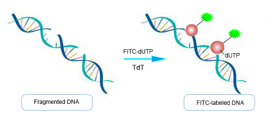
细胞凋亡晚期,染色体DNA双链断裂或单链断裂产生大量的粘性3-OH末端,在脱氧核糖核苷酸末端转移(TdT)的作用下,将荧光素/酶标记的dUTP结合到DNA的3-末端,从而可通过检测荧光完成对细胞凋亡的检测,这类方法称为脱氧核糖核苷酸末端转移酶介导的缺口末端标记法(Terminal-Deoxynucleotidyl Transferase Mediated Nick End Labeling,TUNEL),原理见图1。

图1:脱氧核糖核苷酸末端转移酶介导的缺口末端标记(TUNEL)原理图
产品特点:
适用范围广:可用于石蜡包埋组织切片、冰冻组织切片、培养的细胞和从组织中分离的细胞。
检测灵敏度高:可检测出极少量的凋亡细胞。
背景干扰小:信噪比高。
观察多样性:荧光显微镜观察,流式检测。
数据展示:
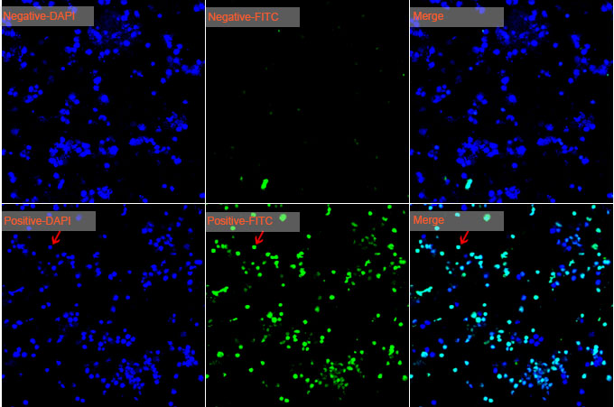
1、小鼠前脂肪细胞(3T3-L)Tunel检测

图2:小鼠前脂肪细胞3T3-L凋亡检测。第一行代表阴性对照,第二行代表阳性对照。
检测试剂:Cat No. 40306 TUNEL Apoptosis Detection Kit (FITC)
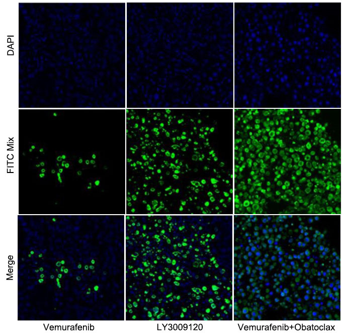
2、石蜡切片样本的Tunel检测

图3:在皮下移植瘤模型中,单独使用LY3009120或联合使用Vemurafenib和Obatoclax可以有效延缓甲状腺癌。IF=8.8
样本类型:石蜡包埋的肿瘤组织(取自裸鼠)。检测试剂:Cat No. 40307 TUNEL Apoptosis Detection Kit (Alexa Fluor 488)。
3、细胞爬片的Tunel检测

图4:硫酸锌对不同分组的内皮细胞凋亡的影响
样本类型:HUVEC(人脐静脉内皮细胞)。检测试剂:Cat No. 40308 TUNEL Apoptosis Detection Kit (Alexa Fluor 640)
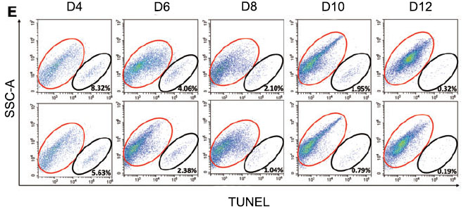
4、流式细胞术检测细胞凋亡

图5:PA处理可以有效降低重编程(reprogramming)过程中凋亡的发生。IF=3.7
样本类型:iPS Cells。检测试剂:Cat No. 40307 TUNEL Apoptosis Detection Kit (Alexa Fluor 488)。
文献引用:
[1] Li C, Wang Q, Gu X, et al. Porous Se@ SiO2 nanocomposite promotes migration and osteogenic differentiation of rat bone marrow mesenchymal stem cell to accelerate bone fracture healing in a rat model[J]. International Journal of Nanomedicine, 2019, 14: 3845. (IF 4.76)
[2] Miao T, Qian L, Yu F, et al. Protective effects of hydroxysafflor yellow an on high oxidized low density lipoprotein induced human coronary artery endothelial cells injuries[J]. Development, 2019, 22: 581-589.(IF 6.208 )
[3] Wen Y, Liu G, Zhang Y, et al. MicroRNA-205 is associated with diabetes mellitus‐induced erectile dysfunction via down-regulating the androgen receptor[J]. Journal of cellular and molecular medicine, 2019, 23(5): 3257-3270.(IF 4.41)
[4] Xu T, Ding W, Ao X, et al. ARC regulates programmed necrosis and myocardial ischemia/reperfusion injury through the inhibition of mPTP opening[J]. Redox Biology, 2019, 20: 414-426.(IF 8.37)
[5] Li P, Hao L, Guo Y Y, et al. Chloroquine inhibits autophagy and deteriorates the mitochondrial dysfunction and apoptosis in hypoxic rat neurons[J]. Life Sciences, 2018, 202: 70-77.(IF 3.40)
[6] Chen H, Guan B, Chen X, et al. Baicalin attenuates blood-brain barrier disruption and hemorrhagic transformation and improves neurological outcome in ischemic stroke rats with delayed t-PA treatment: involvement of ONOO−-MMP-9 pathway[J]. Translational Stroke Research, 2018, 9(5): 515-529.(IF 4.87)
[7] Liu Q, Qian Y, Li P, et al. The combined therapeutic effects of 131iodine-labeled multifunctional copper sulfide-loaded microspheres in treating breast cancer[J]. Acta Pharmaceutica Sinica B, 2018, 8(3): 371-380.(IF 6.88)
[8] Han Y Q, Ming S L, Wu H T, et al. Myostatin knockout induces apoptosis in human cervical cancer cells via elevated reactive oxygen species generation[J]. Redox Biology, 2018, 19: 412-428.(IF 8.37)
[9] Wang S, Xu Y, Weng Y, et al. Astilbin ameliorates cisplatin-induced nephrotoxicity through reducing oxidative stress and inflammation[J]. Food and Chemical Toxicology, 2018, 114: 227-236.(IF 3.78)
[10] Liu L, Pang X L, Shang W J, et al. Over-expressed microRNA-181a reduces glomerular sclerosis and renal tubular epithelial injury in rats with chronic kidney disease via down-regulation of the TLR/NF-κB pathway by binding to CRY1[J]. Molecular Medicine, 2018, 24(1): 49.(IF 3.46)
[11] Li G, Yin Q, Ji H, et al. A study on screening and antitumor effect of CD55-specific ligand peptide in cervical cancer cells[J]. Drug Design, Development and Therapy, 2018, 12: 3899.(IF 3.27)
[12] Qian Y, Wang Y, Jia F, et al. Tumor-microenvironment controlled nanomicelles with AIE property for boosting cancer therapy and apoptosis monitoring[J]. Biomaterials, 2019, 188: 96-106.(IF 9.85)
[13] Li Z, Li D, Li Q, et al. In situ low-immunogenic albumin-conjugating-corona guiding nanoparticles for tumor-targeting chemotherapy[J]. Biomaterials Science, 2018, 6(10): 2681-2693.(IF 5.31)
[14] Liu Y, Zhi X, Hou W, et al. Gd3+-Ion-induced carbon-dots self-assembly aggregates loaded with a photosensitizer for enhanced fluorescence/MRI dual imaging and antitumor therapy[J]. Nanoscale, 2018, 10(40): 19052-19063.(IF 7.17)
[15] Liu Q, Qian Y, Li P, et al. 131 I-labeled copper sulfide-loaded microspheres to treat hepatic tumors via hepatic artery embolization[J]. Theranostics, 2018, 8(3): 785.(IF 8.12 )
常见问题:
Q:出现非特异性荧光标记?
1)组织/细胞本身核酶或聚合酶活性水平较高,易导致出现非特异性的荧光标记,例如平滑肌细胞。解决方法是,取细胞或组织后立即固定并且要充分固定,以阻止这些酶导致假阳性。
2)使用了不适当的固定液,例如一些酸性固定液,导致出现假阳性。建议采用新鲜配置的4%中性对聚甲醛固定液。
3)TUNEL检测反应时间过长,细胞或组织表面不能保持湿润,也可能出现非特异性荧光。注意控制反应时间,并确保TUNEL检测反应液能很好地覆盖样品。
Q:荧光背景很高?
1)高速分裂和增殖的细胞,转录水平高,有时也会出现细胞核中的DNA断裂。
2)在激发光下长时间的暴露,导致假阳性的出现。
Q:标记效率低?
1)使用乙醇或甲醇固定会导致标记的效率较低。
2)固定时间过长,导致交联程度过高。此时宜减少固定时间。
3)组织切片过厚,使得固定效果不理想,最好控制在10 μm以内。
4)荧光淬灭。Fluorescence在普通光照10min就会严重淬灭。解决方法是需注意避光操作。
相关产品:
|
产品名称 |
货号 |
规格 |
|
40311ES20/50/60 |
20T/50T/100T |
|
|
40302ES20/50/60 |
20T/50T/100T |
|
|
40310ES20/50/60 |
20T/50T/100T |
|
|
40303ES20/50/60 |
20T/50T/100T |
|
|
40313ES60 |
100T |
|
|
40305ES20/50/60 |
20T/50T/100T |
|
|
40304ES20/50/60 |
20T/50T/100T |
|
|
40312ES20/50/60 |
20T/50T/100T |
|
|
40306ES20/50/60 |
20T/50T/100T |
|
|
40307ES20/50/60 |
20T/50T/100T |
|
|
40308ES20/50/60 |
20T/50T/100T |
更多产品敬请详询400-6111-883
HB190829
